Importance
The chapter on "control and coordination" is assigned a weightage of \(3\) to \(7\) marks, highlighting its significance in the overall curriculum. Understanding this chapter will enhance the general view of movement in plants, animals, controlled movements, and coordination of movements.
- Section A or B (\(1\) mark or \(2\) marks) - One question
- Section C or D (\(3\) marks or \(4\) marks) - One question
Learning outcomes
-
Movement and nervous system overview: Students describe how animals and humans show controlled movement using the nervous system.
-
Nervous system structure and function: Students identify CNS/PNS parts and explain brain, neuron structure, and impulse transmission.
-
Coordination and reflexes: Students describe nerve impulse conduction, neuromuscular coordination, and reflex arcs.
Detection of Stimuli and role of receptors
Our body detects changes in the environment through receptors located in sense organs such as the skin, tongue, ear, nose and eyes.
This impulse travels from the dendrite to the cell body, moves along the axon, and finally crosses a synapse with the help of chemicals to reach the next neuron or a muscle cell.
The receptors convert a stimulus like heat, taste or smell into a chemical reaction, which then generates an electrical impulse.
Exam tips
-
Always mention that receptors convert stimulus → chemical change → electrical impulse
-
Write examples : Gustatory = taste, olfactory = smell
-
In neuron diagram questions, label cyton, dendrite, axon, synapse clearly
-
Use the term “specialised nerve endings” for receptors
Exam questions:
Structure of neuron
A neuron is the basic unit of the nervous system that receives and transmits impulses. It consists of a cell body, dendrites and a long axon ending in synaptic terminals.
Dendrites receive signals, the cell body processes them, and the axon carries impulses away. Neurons may be apolar, unipolar, bipolar or multipolar, and their axons can be myelinated or non-myelinated.
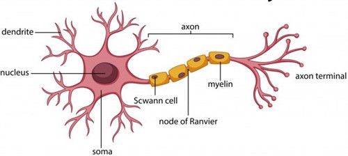
Neuron anatomy
Exam Tips
-
Always state the impulse direction: dendrite → cell body → axon.
-
Myelinated fibres have nodes of Ranvier; non-myelinated do not.
-
Bipolar neurons → retina; multipolar neurons → brain cortex (frequently asked).
Transmission of nerve impulses
A neuron carries information through a fixed path - from its dendritic tip to the axon terminal. At the axon end, the electrical impulse triggers chemicals that pass across the synapse and start a new impulse in the next neuron. This organised network of neurons allows quick and coordinated communication throughout the body.
The action of sensory and motor neuron
Exam Tips
-
Remember the sequence: dendrite → cell body → axon → synapse.
-
Use the phrase “chemicals help the impulse cross the synapse.”
-
In short answers, mention “electrical impulse becomes chemical signal at synapse.”
-
Do not confuse direction of impulse - it always travels one-way.
Exam questions: Structure of neuron
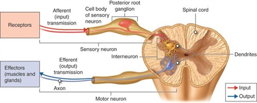
Reflex actions and reflex arc
Reflex actions are quick, automatic responses that occur without conscious thought, like pulling your hand away from a flame.
Instead of waiting for the brain to think and respond, the input nerve and output nerve are connected through a reflex arc in the spinal cord. This shortcut saves time and protects the body from danger, allowing rapid action even before the brain fully processes the signal.

Process of reflex action
Exam Tips
-
Define reflex clearly: “quick, automatic, involuntary response.”
-
Write that the reflex arc is formed in the spinal cord, not the brain.
-
Use examples like touching a hot object or knee-jerk reaction.
-
Differentiate reflex action vs voluntary action in one line if asked.
Exam question: Reflex action
Functions of the human brain
The brain is the main coordinating centre, supported by the spinal cord. The fore-brain handles thinking, decision-making and voluntary actions, and has specialised centres for sensory reception and hunger.
The mid-brain and hind-brain control involuntary activities like heartbeat, breathing and blood pressure. The cerebellum, in the hind-brain, maintains posture, balance, and precision in voluntary movements.
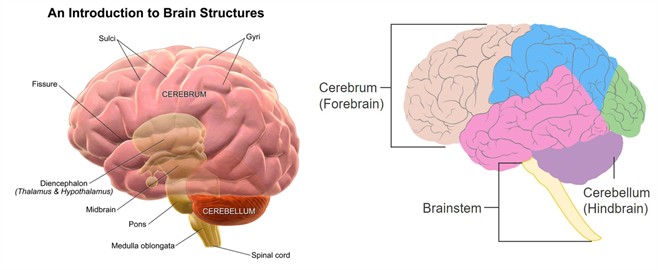
Pictures showing the regions of the brain
Exam tips
-
Fore-brain = Thinking, memory, voluntary actions.
-
Mid-brain + hind-brain = Involuntary actions
-
Cerebellum = Posture + balance + precision
-
Medulla = Heartbeat, breathing, vomiting (frequently asked in exams)
Exam questions
Protection of the brain and Spinal cord
Because the brain is delicate and vital, it is protected inside a bony skull (cranium), and cushioned by fluid-filled membranes (CSF) that absorb shocks. The vertebral column similarly protects the spinal cord and supports the entire neural pathway.
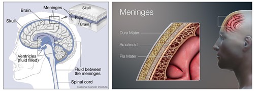
The location of cranial meninges and cerebrospinal fluid
Exam tips
-
Skull protects brain; vertebral column protects spinal cord.
-
Cerebrospinal fluid (CSF) provides cushioning/shock absorption.
-
Use the phrase “bony covering + fluid protection”.
-
Diagram-based questions often ask location and protection — label neatly.
How nervous tissue causes action
Muscle movement occurs when a nerve impulse reaches a muscle fibre, triggering special proteins inside the muscle cell to slide and change shape. This shortens the muscle, producing movement.
Voluntary muscles work under conscious control (like walking), while involuntary muscles function without conscious effort (like heartbeat and digestion).
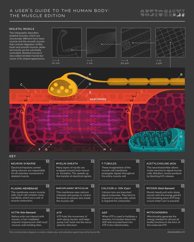
Contraction and relaxation of muscles due to neuron signal
Exam tips
-
Muscle contraction happens due to sliding of proteins inside muscle fibre.
-
Nerve impulse triggers contraction - link both in answers.
-
Voluntary vs involuntary muscles - give one example each.
-
Mention that voluntary muscles respond to brain; involuntary respond automatically.