Although living organisms appear different, all are composed of cells that can be seen only with a microscope, which opened the hidden world of cells and microorganisms explored in this chapter.
Activity
A round-bottom flask filled with water, along with a water droplet, acts like a magnifying glass, making letters and small organisms appear larger and clearer.

Magnifying property of a water droplet
Discovery of Cells
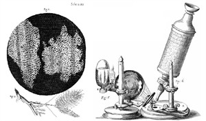
Robert Hooke's microscope
The invention of the microscope helped humans see tiny structures invisible to the naked eye.
In \(1665\), Robert Hooke observed thin slices of cork, and named the small compartments cells, and published a book called Micrographia.
Antonie van Leeuwenhoek later observed living cells, such as bacteria and blood cells, and is known as the Father of Microbiology.
What Is a Cell?
All living things consist of small units called cells; cells are the smallest structural and functional unit of an organism.
-
All plants, animals, and microorganisms are made of one or more cells.
-
Scientists observe cells using a microscope.
Activity:
Aim: To observe the structure of plant cells by preparing a temporary mount of onion peel under a microscope.
Materials required: Onion bulb, forceps, safranin stain, glycerin, glass slide, coverslip, microscope.
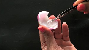
Peeling onion skin
Procedure: A thin onion peel is removed, stained with safranin, washed, mounted on a slide with glycerin, covered with a coverslip, and observed under a microscope.
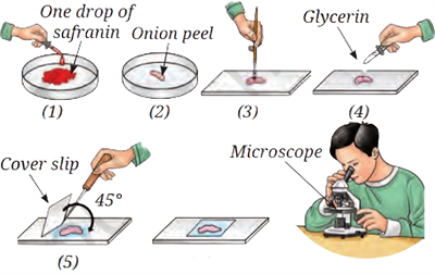
Procedure for the onion peel experiment
Observation: Rectangular, closely packed structures are seen clearly under the microscope.
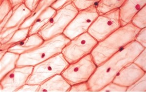
Onion peel skin under the microscope
Conclusion: These structures are onion cells, indicating that plant bodies are composed of many cells, and a microscope is needed to observe them.
Similarly, done for animal cells.
Activity:
Aim: To observe animal cells by preparing a temporary mount of human cheek cells.
The following supplies are needed: a coverslip, glycerin, a methylene blue stain, a glass slide, a clean toothpick, and a microscope.

Scraping of Cheek cells
Method: Cheek cells are carefully scraped, put on a water-filled slide, stained with methylene blue, mounted with glycerin, covered with a coverslip, and examined under a microscope.
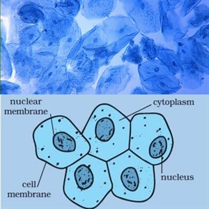
Microscopic and marked human cheek cells
Observation: A microscope reveals polygon-shaped cells with a clear nucleus.
In conclusion, these cheek cells show that cells compose animal bodies.
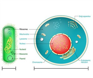
Basic Parts of a Cell
All living things possess cells. Cells are the building blocks of life. The shape, size and number of cells vary across different organisms.
A cell has three significant components.
A cell has three significant components.
1. Cell membrane
2. Cytoplasm
3. Nucleus
2. Cytoplasm
3. Nucleus
In a plant cell, a cell wall surrounds the cell membrane.
Cell Organelles and Their Functions
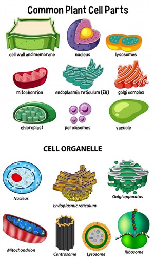
Plant and animal cell organelles
| Cell Organelle | Function |
|---|---|
| Cell membrane | Encloses the cell, separates it from other cells, and controls the entry and exit of substances. |
| Cytoplasm | Contains cell components and is the site where most life processes occur. |
| Nucleus | Controls all cell activities and regulates growth. |
| Cell wall | Provides rigidity, strength, and support to plant cells. |
| Vacuole | Stores food, water, and waste products and helps maintain the cell's shape. |
| Plastids (Chloroplasts) | Contain chlorophyll and help in photosynthesis in green plant cells. |
| Plastids (Leucoplasts) | Help store food substances in plant cells. |
| Plastids (Chromoplasts) | Contain coloured pigments other than chlorophyll and give colour to fruits and flowers. |
| Mitochondria | Release energy needed for various cellular activities. |
Differences Between Plant Cell and Animal Cell
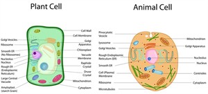
Plant Cell and Animal Cell
| Feature | Plant Cell | Animal Cell |
|---|---|---|
| Cell wall | Present | Absent |
| Cell membrane | Present | Present |
| Chloroplast | Present | Absent |
| Vacuole | Large and permanent | Small or absent |
| Shape | Usually rectangular | Usually round or irregular |
| Mode of nutrition | Makes its own food | Depends on others for food |
Variation in Shape and Structure of Cells
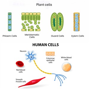
Different types of plant cells and human cells according to their shapes
- Cells have different shapes, and each shape is suited to perform a specific function in the body.
- The cell membrane encloses each cell and maintains its shape. Plant and animal cells usually have permanent shapes.
- Specialised cells, such as nerve cells (long for impulse transmission) and smooth muscle cells (for the movement of organs), show how shape supports function.
Levels of Organisation
The following levels categorise the organisation of living bodies:
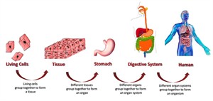
Levels of Organisation
Cell \(\rightarrow\) Tissue \(\rightarrow\) Organ \(\rightarrow\) Organ System \(\rightarrow\) Organism.
The Biggest Cell:
The yolk of an ostrich egg is a single, largest known cell measuring about \(130)\–\(170)\ mm in diameter, protected by a shell and nourished by egg white for its development.
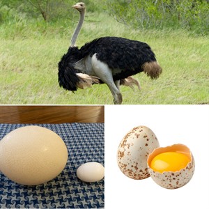
Ostrich egg
Unicellular and Multicellular Organisms
Unicellular and Multicellular
| Feature | Unicellular Organisms | Multicellular Organisms |
|---|---|---|
| Number of cells | Made up of a single cell | Made up of many cells |
| Body organisation | One cell performs all life functions | Different cells perform specialised functions |
| Level of complexity | Simple in structure | Complex in structure |
| Growth | Growth occurs by increase in cell size | Growth occurs by an increase in the number of cells |
| Nucleus | May not have a true nucleus (e.g., bacteria have a nucleoid) | Have a well-defined nucleus in cells |
| Examples | Bacteria, Amoeba, Paramecium, Yeast | Humans, plants, animals, mould |
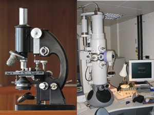
Compound and Electron Microscope