Cell Division: Mitosis, Meiosis, and Tumour Formation
What is Cell Division?
Cell division is the fundamental biological process that enables growth, repair, and development in living organisms. Through this process, a parent cell produces new cells that are essential for:
- Growth and development
- Repair of tissues
- Reproduction (in certain cells and organisms)
If this process were not strictly regulated, errors could lead to serious consequences such as genetic disorders or disease. To prevent this, complex mechanisms ensure that every new cell receives the correct genetic information. The two main types of cell division—mitosis and meiosis—serve distinct functions in the body.
Mitosis (Equational Division):
Mitosis is the process by which a single parent cell divides to form two genetically identical daughter cells, each with the same number of chromosomes as the parent cell. This ensures genetic consistency across body cells.
Where it occurs:
- Somatic (body) cells
- Example: skin, muscles
Stages of Mitosis:
Mitosis occurs in four main stages:
- Prophase – Chromosomes condense and become visible; spindle fibres begin to form, and the nuclear envelope starts to break down.
- Metaphase – Chromosomes align at the centre of the cell along the equatorial plate.
- Anaphase – Sister chromatids separate and move towards opposite poles of the cell.
- Telophase – New nuclear envelopes form around each set of chromosomes, resulting in two distinct nuclei.
- Cytokinesis – The cytoplasm divides, producing two genetically identical daughter cells.
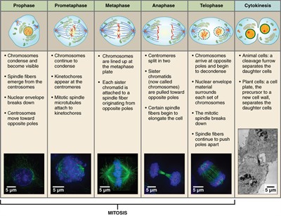
Mitosis
Functions of Mitosis:
- Growth of the body.
- Repair and replacement of damaged cells.
- Maintains chromosome number in somatic (body) cells.
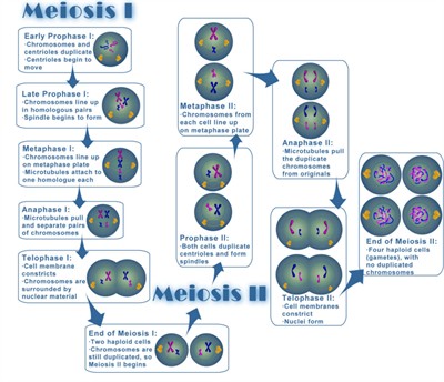
Meiosis (Reduction Division):
Meiosis is a specialised form of cell division in which a single parent cell produces four genetically distinct daughter cells, each with half the number of chromosomes of the original cell. This reduction is essential for maintaining chromosome number during sexual reproduction.
Where it occurs:
- Reproductive organs (testes and ovaries).
- Produces gametes (sperm in males, eggs in females).
Stages of Meiosis:
Meiosis I (Reductional Division):
- Chromosome number reduces from 2n to n
- Homologous chromosomes pair and separate.
- Crossing over occurs in Prophase I
- Produces 2 haploid cells
Stages (Types)
- Prophase I – Synapsis and crossing over (five-sub stages).
- Leptotene – Chromosomes begin to condense and become visible.
- Zygotene – Homologous chromosomes pair (synapsis).
- Pachytene – Crossing over occurs (exchange of genetic material).
- Diplotene – Homologous chromosomes start separating; chiasmata are visible.
- Diakinesis – Chromosomes fully condense; nuclear membrane breaks down.
- Metaphase I – Homologous pairs align at the equator.
- Anaphase I – Homologous chromosomes separate.
- Telophase I – Two haploid cells form.
Meiosis II (Equational Division):
- Similar to mitosis.
- Sister chromatids separate.
- Chromosome number remains the same (n).
- Produces 4 haploid cells.
Stages (Types)
- Prophase II – Chromosomes condense.
- Metaphase II – Chromosomes align at the equator.
- Anaphase II – Sister chromatids separate.
- Telophase II – Four haploid cells form.
Meiosis
Key Features:
- Two successive divisions: Meiosis I (reductional) and Meiosis II (equational).
- Results in four genetically diverse daughter cells.
- Chromosome number is halved (from diploid [2n] to haploid [n]).
Importance:
- Maintains a stable chromosome number across generations in a species.
- Introduces genetic variation through crossing over and independent assortment of chromosomes during meiosis.
Differences Between Mitosis and Meiosis:
|
Properties
|
Mitosis
|
Meiosis
|
|
Type of cells
|
It takes place in somatic cells.
|
It takes place in reproductive cells.
|
|
Number of divisions
|
Single division
|
Two divisions
|
|
Growth and formation
|
Involved in growth and repair; occurs throughout life
|
Involved in gamete formation during reproductive age.
|
|
Synapsis of homologous chromosomes
|
It does not occur
|
In prophase I, crossing over occurs between the non-sister chromatids of chromosomes.
|
|
Genetic composition
|
Genetically identical daughter cells are formed.
|
Daughter cells are genetically different due to recombination and independent assortment.
|
|
Number of daughter cells
|
Two diploid daughter cells are formed.
|
Four haploid daughter cells are formed.
|
|
Chromosome number
|
The chromosome number present in the daughter cell is similar to the parent cell (\(2n\)).
|
The chromosome number present in the daughter cell is just half (\(n\)) of the parent cell.
|
Uncontrolled Cell Division and Tumour Formation:
Normal Cell Division:
Cell division is normally controlled by:
- Cell cycle checkpoints.
- Regulatory proteins (e.g., cyclins).
These controls ensure that cells divide only when needed and help prevent the propagation of damaged or abnormal cells.
What is Uncontrolled Cell Division?
When this regulation fails:
- Cells continue dividing rapidly.
- Damaged or abnormal cells are not removed.
- Leads to the accumulation of abnormal cells.
This process, known as uncontrolled cell proliferation, can lead to serious health issues such as tumour formation and cancer.
What is a Tumour?
A tumour is a mass of abnormal cells that arises when cell division becomes uncontrolled, often due to mutations in genes that regulate the cell cycle.
Types of Tumours:
1. Benign Tumour:
- Non-cancerous
- Localized
- Does not spread
2. Malignant Tumour (Cancer):
- Cancerous
- Invades nearby tissues
- Can spread to other parts (metastasis)
How Tumours Form (Scientific Explanation):
- DNA damage or mutation occurs.
- Cell cycle control fails.
- Cells ignore signals to stop dividing.
- Continuous mitosis leads to tumour formation.
Uncontrolled cell division is a primary cause of cancer development, as it allows abnormal cells to multiply and invade healthy tissue.