The structures that perform specialised functions within themselves are called cell organelles. These cell organelles are the main characteristic that differentiates eukaryotic cells from prokaryotic cells.
The study of cell structure and function of plant and animal cells is known as cell biology.
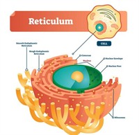
Endoplasmic reticulum:
It is a 3D interconnected network of membrane-lined channels, sheets, and tubules extending from the outer nuclear membrane throughout the cytoplasm.
Endoplasmic reticulum
Parts of ER:
1. Rough endoplasmic reticulum: Appears rough due to ribosomes on its surface and is involved in protein synthesis; mainly composed of cisternae.
2. Smooth endoplasmic reticulum: Lacks ribosomes, appears smooth, and is involved in lipid and steroid hormone synthesis; made up of vesicles and tubules.
Function of endoplasmic reticulum:
- RER synthesises proteins, and SER forms lipids for membrane biogenesis.
- It aids in the intercellular transport of proteins.
- It provides a structural framework and mechanical support.
- SER helps detoxify drugs in liver cells.
- ER produces proteins and lipids that function as enzymes and hormones.
Golgi apparatus:
Materials synthesised in the endoplasmic reticulum have to be transferred to different parts of the cell. A specific cell organelle that performs this transport of materials is golgi apparatus.
The Golgi apparatus is a cell organelle first described by Camillo Golgi.
Camillo Golgi, who studied the nervous system, developed the “Black Reaction,” a silver nitrate staining method that revealed fine nerve cell structures. He received the \(1906\) Nobel Prize with Santiago Ramón y Cajal for discoveries on the nervous system’s structure.
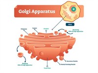
Structure of the Golgi apparatus:
The Golgi apparatus is a stack of membrane-bound, fluid-filled sacs called cisternae that arise from the smooth ER. In plant cells, it is known as the dictyosomes and forms a complex cellular membrane system.
Golgi apparatus structure
The Golgi apparatus is called as the traffic police of the cell because it acts as a way station or assembly area for the storage, processing, and packaging of various cellular secretions.
Functions of the Golgi Apparatus:
- The Golgi apparatus transports, packages, and dispatches materials synthesised near the ER.
- It stores, modifies, and packages products in vesicles.
- It helps form lysosomes.
- It aids in the synthesis of the cell wall and the plasma membrane.
The cis face of the Golgi apparatus receives vesicles from the ER, while the trans face releases processed vesicles.
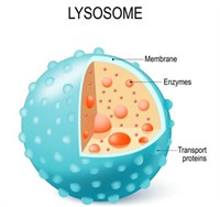
Lysosomes:
Lysosomes are membrane-bound sacs that contain hydrolytic enzymes produced by the RER and function in the disposal of waste. They are present in animal cells but absent in plant cells.
Functions of Lysosomes:
- Lysosomes digest foreign materials and worn-out organelles, serving as the cell’s waste-disposal system.
- Their enzymes break complex substances into simpler ones.
- During starvation, they perform autophagy to release energy.
- Called “suicidal bags” as their rupture can digest the entire cell.
Mitochondria:
The term “mitochondrion” comes from Greek words meaning thread and granule. It is a double-membraned, rod-shaped organelle \(0.5–1.0 µm\) found in both plant and animal cells.
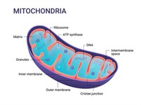
Structure of mitochondrion:
Mitochondria have an outer membrane with porins and an inner folded membrane (cristae) with F1 particles for energy production. The matrix contains enzymes, DNA, and ribosomes essential for ATP synthesis.
Structure of a mitochondria
Functions of Mitochondria:
-
Mitochondria are the cell’s powerhouse, producing ATP through cellular respiration.
-
ATP breakdown releases energy for metabolic activities.
-
They are semiautonomous with their own DNA and ribosomes.
-
They supply intermediates for the synthesis of fatty acids, steroids, and amino acids.
Plastids:
Plastids, found only in plant cells, give plants their colour and enable photosynthesis. The term was coined by Ernst Haeckel in \(1866\). Plastids are of three main types depending on their colour.
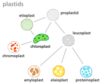
1. Chromoplasts: Colored plastids containing carotenoids, giving red, orange, or yellow hues to flowers and fruits.
2. Leucoplasts: Colourless plastids in storage cells that store food, amyloplasts (starch), aleuroplasts (protein), and elaioplasts (oil/fat).
3. Chloroplast: Green plastids with chlorophyll and other pigments, mainly in leaves, responsible for photosynthesis.
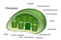
Structure: Plastids vary in shape and contain a semi-fluid stroma, which contains DNA, ribosomes, and enzymes. Their grana are stacks of thylakoids, each containing chlorophyll that captures sunlight for photosynthesis.
Function: Chlorophyll traps the solar energy, which is used for manufacturing food. These are essential for photosynthesis. So, chloroplasts are called the "kitchen of the cells".
Vacuoles:
Water, sugars, minerals, and amino acids are essential to the cell. In plants, these materials are stored in the cell organelle called the vacuole.
In animal cells, vacuoles are small, but in plants, the vacuoles occupy \(50\) to \(90\) of the volume of the cell.
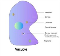
Structure: The vacuole is a large, central organelle bounded by a selectively permeable tonoplast that occupies most of the cell’s volume.
Components: It contains cell sap which is made of water, sugars, minerals, amino acids, and proteins essential for plant cell function.
Function:
- Vacuoles help to provide turgidity and rigidity to the cell.
- Vacuoles act as a storehouse for water-soluble pigments and waste products. It also stores useful minerals and salts.
- The sap vacuole maintains proper osmotic pressure in the cell for its turgidity and absorption of water.
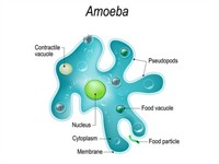
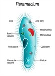
Unicellular organisms like amoebas have food vacuoles; they contain digestive enzymes. With these enzymes , nutrients are digested, and digested food items are further stored in it. Contractile vacuoles (present in paramecia) play a key role in expelling excess water and soil wastes.
Ribosomes:
The proteins are essential for the structure, function, and regulation of the body's tissues and organs, and they perform most of their work in cells. Ribosomes carry out protein synthesis inside a cell.
Cell division:
Cell division is a process of the formation of new cells from pre-existing cells.
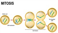
1. Mitosis or Mitotic cell division:
It is the kind of cell division where a cell divides to produce two genetically identical daughter cells from the dividing cells of the mother cell.
Mitosis occurs in almost all the body's somatic cells, which includes those of the eyes, skin, hair, and muscles. It is also known as equational division.
Mitosis division
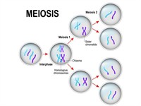
2. Meiosis or Meiotic cell division
This kind of cell division forms the gametes. The male (sperm) and female (egg) gametes, both are haploid, unite to form a zygote (a diploid cell) after fertilization, which gives rise to offspring (young ones). When a cell divides by meiosis, it forms four new cells instead of two.
This cell division occurs in the sexually reproducing cells.
Meiosis division