Transplantation is needed when an organ fails due to disease or injury and involves surgically transferring organs like the kidney, heart, liver, or cornea from a living or deceased (brain-dead) donor.
Why is the heart divided into chambers?
The separation of the heart is important to avoid the mixing of oxygenated and deoxygenated blood. The separation also allows for an efficient supply of oxygen to the body.
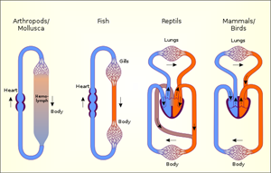
Amphibians and reptiles have a \(3\) chambered heart that tolerates some mixing of oxygenated and deoxygenated blood. In other vertebrates, blood goes through the heart twice during each cycle of passage through the body. The above is called double circulation.
Circulation of blood in different organisms
1. Fishes have a \(2\)-chambered heart (one atrium, one ventricle). Blood passes through the heart only once, showing single circulation.
2. Amphibians and most reptiles have a \(3\)-chambered heart (two atria, one ventricle). Partial mixing of oxygenated and deoxygenated blood occurs.
3. Crocodiles, birds, and mammals have a \(4\)-chambered heart (two atria, two ventricles). There is no mixing of blood, ensuring double circulation.
Circulation in different organisms
Blood and its vessels
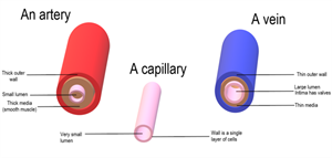
Arteries:
- Arteries carry oxygenated blood from the heart to the different parts of the body.
- They have thick, elastic walls as blood emerges from the heart under high pressure.
- Arteries are narrow in size as the blood flows quickly through them.
Veins:
- Veins collect the deoxygenated blood from different organs and pour it into the heart.
- Since they collect blood from the body parts, they are wider.
- Veins do not have thick walls, as the blood is not under pressure. They have valves that prevent the backward flow of blood.
Important!
The pulmonary artery is an exception which carries deoxygenated blood from the right ventricle to the lungs. The pulmonary vein is an exception and carries oxygenated blood from the lungs to the left atrium.
Capillaries:
When an artery reaches a tissue or an organ, it divides into smaller vessels to contact all the individual cells. Capillaries are the smallest vessels which are one cell thick. The thin wall of capillaries allows the exchange of substances between the blood and the surrounding cells.
Blood vessels
What is meant by Blood pressure?
The pressure exerted by the flow of blood on the thick and elastic walls of the arteries is called blood pressure.
The pressure is greater in the arteries than in the veins. The pressure of blood in the artery during ventricular systole is known as systolic pressure. The pressure in the artery during ventricular diastole is known as diastolic pressure.
Healthcare providers measure blood pressure using an instrument called a sphygmomanometer. In a normal person, the systolic pressure is \(120\ mmHg\) and the diastolic pressure is \(80\ mmHg\). Doctors express it as \(120/80\).
Hypertension is a sustained increase in blood pressure. It occurs due to arteriole constriction, increasing blood flow resistance and may cause artery rupture and internal bleeding.
Blood clotting: When an injury damages a blood vessel, bleeding starts; however, blood clotting or blood coagulation. To prevent this, the blood contains platelets that circulate throughout the body and help stop bleeding.
Human excretory system
The body removes harmful metabolic wastes through a process called excretion.
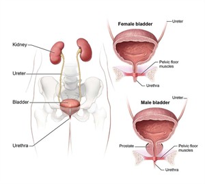
The human excretory system removes wastes via skin (sweat), lungs \(CO_2\), and the urinary system (urine). It consists of kidneys, ureters, urinary bladder, and urethra, which help eliminate excess water, salts, and urea.
Human excretory system
- Kidneys: The kidneys are bean-shaped organs on either side of the backbone that filter blood to form urine. Each kidney contains about \(10\ \)lakh nephrons, the structural and functional units of the kidney.
- Ureters: The ureter of each kidney is a narrow, tubular structure that opens into the urinary bladder. Ureters carry urine from the kidneys to the urinary bladder.
- Urinary bladder: The urinary bladder is the reservoir of urine that stores urine temporarily.
- Urethra: The urethra is a canal-like structure that opens into the exterior through the urethral orifice. Urine produced in the kidneys passes through the ureters into the urinary bladder. The bladder stores the urine until the body releases it through the urethra.
Nephrons - The Unit of the Excretory System
-
Blood enters the kidney from the aorta through the renal artery, which divides into smaller vessels forming afferent arterioles.
-
Each afferent arteriole enters Bowman’s capsule and forms a capillary network called the glomerulus.
-
The glomerulus and Bowman’s capsule together form the renal corpuscle, which filters blood and removes wastes.
-
The filtrate passes through the proximal convoluted tubule, then the loop of Henle.
-
It then enters the distal convoluted tubule and finally the collecting duct, which carries urine to the ureter.
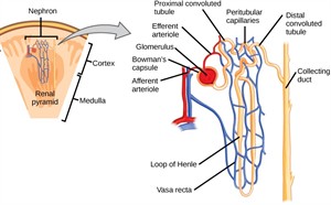
Structure of a nephron
How is urine produced in the kidneys?
Waste is excreted primarily in the form of urine in humans. The kidneys produce urine as they filter waste products from the blood. The nitrogenous metabolic wastes, such as urea or uric acid, are removed from the blood in the kidneys. The nephrons help in the filtration of these wastes from the blood.
The urine composition includes approximately \(95%\) water and \(5%\) solid wastes.
Artificial kidney or Hemodialysis
Kidney damage can cause waste products to accumulate, leading to kidney failure; however, doctors can treat this condition with an artificial kidney. An artificial kidney removes nitrogenous wastes by dialysis using semi-permeable tubes.
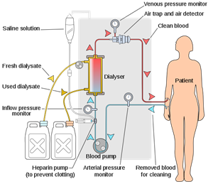
Haemodialysis
Organ Donation and Transplantation.
Organ donation is giving an organ to a person with organ failure, with consent, regardless of age or gender, and it can save lives.