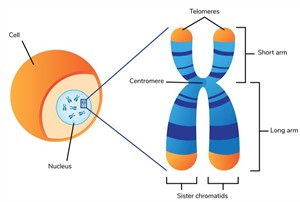
The nucleus of each cell contains thin thread-like structures called chromosomes. Waldeyer first coined the term 'chromosomes' in \(1888\).
Except for RBCs, chromosomes are found in the nucleus of all body cells. Red blood cells have no nucleus and therefore do not contain chromosomes.
Chromosomes contain hereditary information and carry genetic material. Chromosomes are folds of chromatin fibres packed with DNA and proteins that form the genetic material.
Sister chromatids: Two identical chromosome strands joined at the centromere, composed of chromonema (with chromomeres) and made of DNA, RNA, and proteins for structural support.
Chromosome regions:
1. Primary constriction (Centromere): Region where sister chromatids are joined, and spindle fibres attach during cell division.
2. Secondary constriction: Narrow region (nucleolar organiser) involved in nucleolus formation.
3. Telomere: Terminal end of a chromosome that provides stability and prevents fusion with other chromosomes.
4. Satellite: Small knob-like extension at the chromosome end; chromosomes bearing it are called sat-chromosomes.
The structure of the chromosome
Based on the position of the centromere
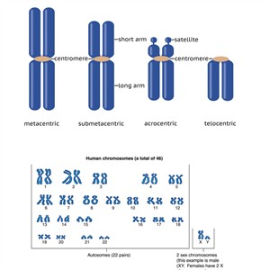
Chromosomes are classified by centromere position and type.
1. Telocentric: Centromere at the terminal end, forming a rod-shaped chromosome.
2. Acrocentric: Centromere near one end, producing one short and one long arm (rod-shaped).
3. Submetacentric: Centromere slightly off-centre, forming unequal arms (J or L shaped).
4. Metacentric: Centromere at the centre, forming two equal arms (V-shaped).
2. Acrocentric: Centromere near one end, producing one short and one long arm (rod-shaped).
3. Submetacentric: Centromere slightly off-centre, forming unequal arms (J or L shaped).
4. Metacentric: Centromere at the centre, forming two equal arms (V-shaped).
Types of chromosomes
1. Autosomes: Chromosomes that control body (somatic) traits, present in equal numbers in both males and females.2. Allosomes (Sex chromosomes): Chromosomes that determine sex, namely X and Y chromosomes.
The number, size, and shape of chromosomes in an organism's cell nucleus are called a karyotype.
DNA molecule
DNA is a large molecule with several nucleotides in it. Thus, it is also known as a polynucleotide.
-
Each Nucleotide consists of deoxyribose sugar, a nitrogenous base (purine or pyrimidine), and a phosphate group (\(H₃PO₄\)).
-
DNA bases are of two types: Purines (Adenine and Guanine) and Pyrimidines (Cytosine and Thymine).
-
\(Nitrogen\ base + Sugar = Nucleoside\); \(Nucleoside + Phosphate = Nucleotide\).
Watson and Crick model of DNA
1. DNA consists of two antiparallel polynucleotide strands forming a double helix with a sugar-phosphate backbone linked by phosphodiester bonds.
2. Bases show complementary pairing \(A = T\) (2 H-bonds) and \(G ≡ C\) (3 H-bonds) following Chargaff's rule; in RNA, uracil (U) replaces thymine.
3. Hydrogen bonds stabilise DNA, measure \(20 Å\) in width, \(34 Å\) per turn, and have \(10\ \)base pairs per helical turn.
2. Bases show complementary pairing \(A = T\) (2 H-bonds) and \(G ≡ C\) (3 H-bonds) following Chargaff's rule; in RNA, uracil (U) replaces thymine.
3. Hydrogen bonds stabilise DNA, measure \(20 Å\) in width, \(34 Å\) per turn, and have \(10\ \)base pairs per helical turn.
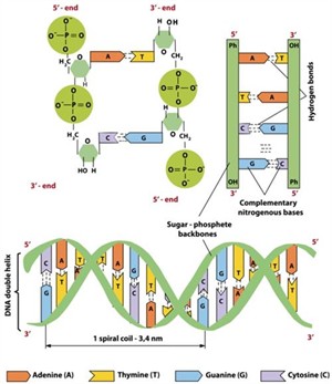
The structure of DNA
DNA replication
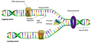
1. Origin of replication: The specific DNA site where replication begins and the strands separate to form a replication fork.
2. Unwinding of the DNA molecule: Helicase separates the two strands, while topoisomerase relieves supercoiling ahead of the fork.
3. Formation of RNA primer: A short RNA primer is synthesised to provide a starting point for DNA synthesis.
4. Synthesis of new strand: DNA polymerase adds nucleotides to form a complementary strand in a\(\ 5′→3′ \)direction.
5. Leading and lagging strands: The leading strand is synthesised continuously, while the lagging strand forms Okazaki fragments joined by ligase.
6. Termination: Replication ends when replication forks meet at the terminus region.
DNA replication
Mutation
When a DNA gene is destroyed or altered so that the genetic message carried by that gene is altered, it is called a mutation. A mutation changes an organism's genetic material (DNA), which can be passed down through generations.
Chromosomal mutation: A sudden change in the structure or number of chromosomes.
Types of mutation
1. Structural changes: Caused by cell division errors leading to deletion, duplication, inversion, or translocation.
2. Numerical changes: Gain or loss of chromosomes, resulting in changes in ploidy.
Ploidy types:
Euploidy: Presence of extra complete chromosome sets (e.g., Triploidy – \(3n\), usually sterile; Tetraploidy –\(\ 4n\), often produces larger plants).
Aneuploidy: Gain or loss of individual chromosomes, such as Monosomy (\(2n−1\)), Trisomy (\(2n+1\)), and Nullisomy (\(2n−2\)).
Euploidy: Presence of extra complete chromosome sets (e.g., Triploidy – \(3n\), usually sterile; Tetraploidy –\(\ 4n\), often produces larger plants).
Aneuploidy: Gain or loss of individual chromosomes, such as Monosomy (\(2n−1\)), Trisomy (\(2n+1\)), and Nullisomy (\(2n−2\)).
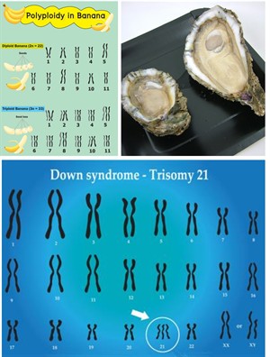
Euploidy - diploid and polyploid banana, diploid and triploid oyster and Down syndrome.
Down syndrome:
A common aneuploid condition (Trisomy \(21\)) was first described by John Langdon Down in \(1866\).
Features: Characterised by intellectual disability, delayed development, weak muscle tone, and vision/hearing problems.
Gene (point) mutation: A change in the nucleotide sequence of a gene due to substitution, insertion, deletion, or inversion of bases, leading to abnormal protein formation.
Sex determination
Females are homogametic (\(XX)\) in this type of sex determination, which means they produce only one type of gamete. Females have two \(X\) chromosomes.
Males are heterogametic (\(XY\)), which means they produce two types of gametes.
In males, \(X\ \)chromosome-containing gametes are referred to as gynosperm, while \(Y\) chromosome-containing gametes are referred to as androsperm.
Males are heterogametic (\(XY\)), which means they produce two types of gametes.
In males, \(X\ \)chromosome-containing gametes are referred to as gynosperm, while \(Y\) chromosome-containing gametes are referred to as androsperm.
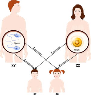
\(XX-XY\) type sex-determination system in humans
Fertilisation of the egg (\(22+X\)) with a sperm (\(22+X\)) results in the birth of a female child (\(44+XX\)).
Fertilisation of the egg (\(22+X\)) with a sperm (\(22+Y\)) results in a male child (\(44+XY\)). The father determines the child's sex.
Other types
1. \(XX–XO\) type: Males are heterogametic (\(XO\)) and females homogametic (\(XX\)); seen in grasshoppers and cockroaches.
2. \(ZW–ZZ\) type: Females are heterogametic (\(ZW\)) and males homogametic (\(ZZ\)); seen in birds and some reptiles.
3.\(\ ZO–ZZ\) type: Females have a single Z chromosome (\(ZO\)), and males have a double Z (\(ZZ\)); seen in butterflies and moths.
2. \(ZW–ZZ\) type: Females are heterogametic (\(ZW\)) and males homogametic (\(ZZ\)); seen in birds and some reptiles.
3.\(\ ZO–ZZ\) type: Females have a single Z chromosome (\(ZO\)), and males have a double Z (\(ZZ\)); seen in butterflies and moths.